
The major distribution of beta receptors is: 1 heart, 2 lungs, 3 adiposeīlebitis. Order of use: Prostaglandins > Beta-blockers > Alpha 2 agonists > Cholinergics > Carbonic anhydrase inhibitors Betaxolol is a relatively selective beta-1 blocker which in most patients is almost as effective as timolol in lowering intraocular pressure, and may be partly.

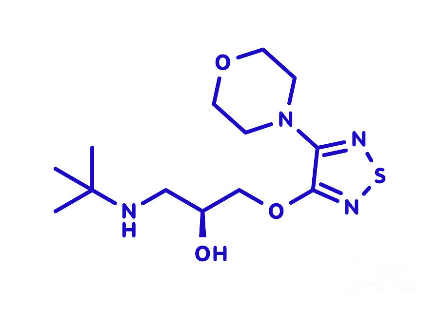
It is a medicine which is used to treat several different medical conditions. Oral ( acetazolamide) is more effective because it also blocks isoenzyme IV Timolol belongs to the group of medicines known as beta-blockers.Inhibit anhydrase isoenzyme II → reduce aqueous production.Cholinergic medications are not used in secondary angle closure because contraction of the ciliary body can cause it to rotate forward, making the anterior chamber even shallowerĬarbonic anhydrase inhibitors (e.g Brinzolamide (any -lamide)).Also causes miosis which relieves pupil block, where the the pupil has fallen down and is obstructing the flow of aqueous.Muscarine agonism leads to ciliary contraction, opening the trabecular meshwork → Increases trabecular outflow.A2 selective blockers are lipophilic, and can cross the blood brain barrier, resulting in respiratory depression in the young.Decrease aqueous production and increase uveoscleral outflow.This means that it doesn’t cause as many side effects Carteolol is a competitive intrinsic agonist which has intrinsic sympathomimetic properties.Betaxolol is a selective beta 1 blocker.Block adrenergic cAMP → reduces aqueous production.Avoid in uveitis as it worsens inflammation Timolol maleate is a beta 1 and beta 2 (non-selective) adrenergic receptor blocking agent that does not have significant intrinsic sympathomimetic, direct myocardial depressant, or local anesthetic (membrane-stabilizing) activity.Analogues of PGF2Alpha → increase uveoscleral outflow.Nasal vessel displacement and haemorrhages at the disc are also signs of glaucomatous changeĭecreased nerve fibre layer (NFL) thickness readings on OCT would make one suspicious for optic nerve damage.In glaucoma, the cup-to-disc ratio is increased.A normal cup-to-disc ratio is around 0.3.In glaucoma, the neuroretinal rim becomes thinner so that it no longer follows the order of the ISN’T rule.Neuroretinal rim thickness follows the ISNT rule of thickness: inferior (thickest), superior, nasal and temporal (thinnest).The centre of the disc is the cup and contains the central retinal vessels The neuroretinal rim is the border of the optic disc and contains the retinal neuronal cells.Glaucomatous diseases processes invariably damage the optic nerve, evidence of this damage can be seen in the optic disc during fundoscopy Note the increased cut-to-disc ratio and asymmetrical thinning of the neuroretinal rim. A healthy optic disc By Ske, CC BY-SA 3.0Īn optic disc in advanced glaucoma.


 0 kommentar(er)
